Looking At German Data For Myocarditis And Other Adverse Effects
We are going to look at two sets of data. The first one is most likely not peer reviewed and the second is peer reviewed or reviewed by other medical professionals.
Again, I am not a medical professional. You should cross examine or talk about this with your doctor or people with a medical degree. It has been a decade since I studied biology.
Professor Arne Burkhardt Explains
This one is in German. So how did I understand this video and his other videos? I used the youtube subtitles. He explained this well and I think he is the first person to come up with this idea so I do believe I should include him on my post.
The process is simple.
He uses various pigments (color) to identify certain cells and tissues. The pigment will highlight lymphocytes which are immune cells or antibodies that destroy other cells the immune system deems foreign or harmful. The pigment also highlights the spike protein. What he concluded was that.
- Vaccine enters the cells, often endothelial cells which are cells that make blood vessels and the heart.
- The vaccine tells the cells to create the spike proteins.
- These spike proteins remain at the surface of the cells.
- The lymphocytes (B-cell and T-cell) recognizes the spike proteins and it launches an immune response to destroy the spike proteins. Thus destroying the cells of the organ the spike proteins are attached to, creating organ damage.
How do we know that the underlying cause is the vaccine and not COVID virus since the virus has the spike protein also. The cells only have the spike proteins and they do not have the other parts of the virus like the nucleotides and so on. If only the spike proteins are present, then we know that it was not caused by the virus.
40 Year Old Woman Biopsy Of The Lower Leg

What is a biopsy? Basically, they removed this tissue on a living person to discover the cause of the underlying disease that the person is experiencing. The blue ones on the left are lymphocytes, in the middle of them are the spike proteins, you can see on the right. The tissue is swollen or inflamed because the lymphocytes are destroying the cells.
Rod-like Substance

There are also these rod-like substances that trigger an autoimmune response. This pic is of the walls of the heart. I’m not sure what it is, the translation is not clear. Some sort of cholesterol needle (google translate) or maybe the metals from the vaccines.

He also said that it is hard to identify what they are. The microscope cannot get a clear picture. When he zooms in, it becomes too close. He does not know where he is anymore.

Here we have an example of what is happening to the blood vessels or the heart. To the left, you have the endothelium which is the thin membrane that lines the inside of the heart and blood vessels. To the right, it is destroyed. I’m not sure whether he is saying when the spike proteins come out, the cell gets destroyed or when the lymphocytes come in, the cell gets destroyed. The bottom line is that the cell gets destroyed.
Where Can The Spike Proteins Go?

They can go anywhere where the blood flows. They can be found in the heart, lungs and brain.

You can see the lymphocytes killing the cells and creating inflammation.

In here you can see the spike proteins attached themselves to the blood vessel walls. There are a lot of them. All clump up together.

Here you can see the spike proteins and the hemorrhage of the coronary artery. Hemorrhage means the blood getting out of the blood vessel.

Here you can see the spike proteins on the heart.
And So On
For the rest of the video, he would just kept providing evidence of spike proteins in different organs like the brain, spleen, lungs, and so on. It is basically the same thing so I just skip it. Basically, he says that when the spike proteins are produced, they don’t float around. They stay where they get produced, attaching themselves to the nearest cells. The immune system destroys the spike protein along with the cell that it is attached to.
So if the vaccine enters the cells of the heart, that cell will produce spike proteins. These spike proteins do not travel. They stay near where they are created attaching themselves to the surrounding cells.
The Video Was Created
The video was created a month ago, I see a date which September 18, 2022 so that maybe the time this was created. This does not seem to be widely accepted. Maybe the evidence hasn’t been peer reviewed or people cannot verify the people who he got the tissues from. I don’t know. I don’t see this research getting cited by other medical professionals yet.
We Go To The Second Set Of Data
Clinical Research In Cardiology

This evidence comes from the Clinical Research in Cardiology. The link to the study can be found here. This has been peer reviewed and the data has been published.
Very Simple Diagram
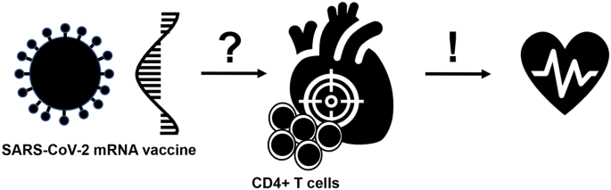
This has a very simple diagram to tell us what is happening. Vaccine enters the cells, creates spike proteins. The vaccine or spike proteins goes to the heart? They don’t know exactly which is why it is a question mark. Heart gets attacked by antibodies and this leads to heart failure. It is so much easier to understand than the one about J&J heparin induced thrombosis.
The Study
The study involves 25 people.
Standardized autopsies were performed on 25 persons who had died unexpectedly and within 20 days after anti-SARS-CoV-2 vaccination.
In four patients who received a mRNA vaccination, we identified acute (epi-)myocarditis without detection of another significant disease or health constellation that may have caused an unexpected death.
Overall, autopsy findings indicated death due to acute arrhythmogenic cardiac failure.
The data was taken by doing autopsies. All autopsies were performed at one of the five University hospital sites (Heidelberg, Tübingen, Freiburg, Ulm, Mannheim) of Baden-Württemberg.
They used the dye formalin to show the lymphocytes and other cells.
For histological evaluation, all tissue samples were fixed in 4% neutral-buffered formalin. At least two full-thickness blocks from the left and right ventricular wall, the interventricular septum, and the papillary muscles were taken for histological evaluation of cardiac pathologies.
The Data
Histological examination showed inflammatory infiltration of the myocardium.
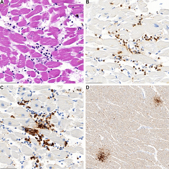
A Lymphocytic aggregates in the interventricular septum of case 1 with associated myocardiocyte destruction. B The infiltrate is predominantly composed of CD3-positive T-lymphocytes and C CD68-positive macrophages. D In lower magnification two foci of CD4-positive lymphocytes are evident (D)
At figure A, you can see the blue cells and the red tissues of the heart. The blue cells are the lymphocytes or antibodies. They basically destroy substances that they deemed foreign. In here, you can see the myocardiocyte destruction which the destruction of the muscular tissue of the heart. The muscles that are responsible for making your heart pump blood or beat.
At figure B and C, we can see the T-lymphocytes and macrophages. Macrophages are a type of white blood cells that kills microorganisms and foreign substances.
At figure D, they zoomed out. We can see that it is very focal. They clump together or concentrated in certain areas of the heart.
Second Image

A Inflammatory focus in the left ventricular wall of case 2. B The infiltrate is predominantly composed of CD68-positive macrophages and C CD3-positive T-lymphocytes with (D) co-expression of CD4
Third Image

A The jab site in the deltoid muscle reveals focal inflammation. The composition is similar to the phenotype of the myocardial infiltrates showing predominantly, B CD3 and C CD4-coexpressing lymphocytes and D interspersed CD68-positive macrophages
This one is from the deltoid muscle where they stick the needle in. The muscle on your shoulder. You can see that what is happening is similar which is the lymphocytes are destroying the cells. What they are saying is that the destruction of the heart muscle cells is similar to the destruction of the deltoid muscle cells so there must be some sort of correlation.
The Data Comes From
Through our autopsy-based approach, we identified five cases of lymphocytic (epi-)myocarditis in persons, who were unexpectedly found dead at home within the first week following mRNA-mediated anti-SARS-CoV-2 immunization. According to the Dallas criteria four samples were classified as definitive myocarditis. In the remaining case, comparable inflammatory infiltration of the epicardium, subepicardial fat and myocardium was found, but myocardial infiltration did not exceed the threshold of the Dallas criteria. All cases showed a consistent phenotype: (A) focal interstitial lymphocytic myocardial infiltration, in three cases accompanied by demonstrable microfocal myocyte destruction. (B) T-cell dominant infiltrate with CD4 positive T-cells outnumbering CD8 positive T-cells by far; (C) frequently associated with T-cell infiltration of epicardium and subepicardial fat tissue revealing a similar immune phenotype (CD4 > > CD8).
It just explains that four of the patients definitely died due to the immune system attacking the heart muscle resulting in heart failure.
This is reflected by the fact that myocarditis is a major cause of sudden and unexpected death in infants, adolescents, and young adults with frequencies ranging from 1 to 14% among the young [18,19,20,21].
They are saying that this data could explain why young people are dying. If you think about it, in 2020, it was the old people who are dying. The young people are fine. Now, all of a sudden, the young people are dying left and right. They are saying that this could be the cause.
How Can People Die From This?
This is from my knowledge. I’m not a medical professional so I could be wrong. You should ask a doctor. There are two ways you could die from this other than hemorrhaging to death or having blood clots. Again, this may not be true. Don’t take this as fact.
The first one when your heart muscles gets damaged, they don’t regenerate. The damaged area is no longer made up of heart muscles. They become scar tissues or connective tissues. Think of it like when your skin gets a scar. The scar no longer looks like normal skin. It becomes scar tissues. It is the same for the heart muscles. They are no longer heart muscles so they stop functioning like one.
The second is that the heart gets electrical impulses. These scar tissues, they do not conduct the same way. It could also happen when the heart muscle is inflamed because it is getting destroyed. The heart does not get the same electrical impulses so it stops working properly. The person most likely needs to go to the hospital asap to get that shocked treatment to make the heart work again.
Again, this is really beyond my expertise. Anyway, an anecdotal experience I read. The person felt like he was going to faint, he had shortness of breath, and good thing they went to the hospital asap because his heart stopped and they had to shock him to get it working again.
How Did They Link This To The Vaccine
In general, a causal link between myocarditis and anti-SARS-CoV-2 vaccination is supported by several considerations: (A) a close temporal relation to vaccination; all cases were found dead within one week after vaccination, (B) absence of any other significant pre-existing heart disease, especially ischaemic heart disease or cardiomyopathy, (C) negative testing for potential myocarditis-causing infectious agents, (D) presence of a peculiar CD4 predominant T-cell infiltrate, suggesting an immune mediated mechanism. The latter criterion is supported by demonstration of a phenotypically identical T-cell infiltrate at the deltoidal injection site in one of the cases. It has to be emphasized, that a comparable (epi-)myocardial infiltration was neither found in any of the other 20 autopsies performed on bodies found dead within 20 days following an anti-SARS-CoV-2 vaccination nor in the age- and sex-matched cohorts from three independent periods from our autopsy-files.
So What Is Missing?
Professor Arne Burkhardt said that the spike proteins attached themselves to the endothelial cells or cells that commonly make up blood vessels and the heart. Unfortunately, they don’t have any evidence of that. They did not have any evidence of the spike proteins being in the heart muscles, blood vessels and so on. I don’t know whether they did not have the proper equipment or it just was not there or the immune system destroyed all of them.
Thus, it seems possible that a molecular mimicry between the spike protein of SARS-CoV-2 and self-antigens may trigger an anti-myocytic immune response in predisposed individuals. Multiple studies of mRNA-vaccines showed robust Receptor-Binding-Domain specific antibodies, T cell and cytokine responses [26]. T cells, especially CD4 + T cells, are the main drivers of heart-specific autoimmunity in myocarditis [27]. A vaccine-induced activation of the immune system in persons with otherwise peripheral tolerance due to regulatory T cells might promote CD4 + effector T cell expansion and myocarditis. Considering that (epi-)myocarditis has not been described following vector-based anti-SARS-CoV-2 immunization yet, it could also be possible that the immune response may be directed against the mRNA or other constituents of the vaccine formula. However, the vaccine against smallpox, based on a vaccinia virus, is reported to cause (epi-)myocarditis in rare cases [2, 3]. Of note, it has been recently reported that intravenous injection of COVID-19 mRNA vaccine is able to induce an acute (epi-) myocarditis in a preclinical model [28]. Interestingly, we recorded inflammatory foci predominantly in the right heart, which may suggest a gradual blood-stream derived dilution effect and based on this finding it is at least tempting to speculate that inadvertent intravascular vaccine injection may be contributive.
Their Conclusion
Finally, we cannot provide a definitive functional proof or a direct causal link between vaccination and myocarditis. Further studies and extended registry are needed to identify persons at risk for this potentially fatal AEFI and may be aided by detailed clinical, serological, and molecular analyses which were beyond the scope of this study. Considering that this fatal adverse event may affect healthy individuals, such registry and surveillance programs may improve early diagnosis, close monitoring, and treatment.

Without the spike proteins, they cannot fully say that it is definitive. What they have is temporal data which basically means that it is based on time. From what I can tell, they are basically saying: These patients all died after 20 days of taking the vaccine, we found the immune system attacking the heart which looks the same as the immune system attacking the jab site. Therefore, there is likelihood that they are the same.
So if you go to the first image (D), you will see that they are concentrated two spots. The study does not know why they are concentrated on those spots but according to Prof. Arne’s statement, they are concentrated on those sites because that is where the vaccine entered the cell, produced spike proteins. The spike proteins attached themselves to the surrounding cells. The immune system destroyed those cells where the spike proteins attached themselves into.
If It Is Proven Causality Between Vaccine And Myocardia
It is going to be crazy if it is proven that vaccines cause myocardia. The society is going to flip. Before the vaccinated do not want to do anything with the unvaccinated. It is going to be the other way around. Is the vaccine contagious? It enters the cell, the cell produces spike proteins. Can this cell be transported through liquid transmission like coughing, sneezing, kissing, sex and so on? Will the other person start producing spike proteins if it does. So many questions.